GUTSCHEIN
Bitte haben Sie einen Moment Geduld, wir legen Ihr Produkt in den Warenkorb.
Diese Produkte könnten Sie auch interessieren:
Bitte haben Sie einen Moment Geduld, wir legen Ihr Produkt in den Warenkorb.
| Reihe | Atlas of Imaging Anatomy |
|---|---|
| Themen | Medizin Medizinische Spezialgebiete Bildgebende Verfahren Radiologie |
| ISBN | 9781626239869 |
| Sprache | Englisch |
| Erscheinungsdatum | 14.10.2026 |
| Größe | 30.5 x 22.9 cm |
| Verlag | Thieme Medical Publishers |
| Lieferzeit | Erscheint am 14.10.2026 |
| Herstellerangaben | Anzeigen Georg Thieme Verlag KG Oswald-Hesse-Straße 50 | DE-70469 Stuttgart info@thieme.de |
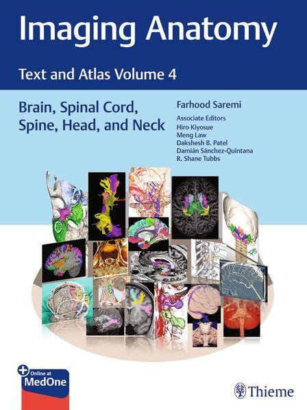
A visually rich radiologic guide to the brain, spinal cord, head, and neck structures
Normal imaging anatomy and variants, including diagnostic and surgical anatomy, are the cornerstones of radiologic knowledge. Imaging Anatomy: Text and Atlas, Volume 4: Brain, Spinal Cord, Spine, Head, and Neck is the fourth in a series of richly illustrated radiologic references edited by distinguished radiologist Farhood Saremi. The atlas is coedited by esteemed colleagues Hiro Kiyosue, Dakshesh Patel, Damian Sanchez-Quintana, and R. Shane Tubbs with contributions from an impressive cadre of international authors. Succinctly written text and superb images provide readers with a virtual, user-friendly dissection experience.
This exquisitely crafted atlas combines fundamental core anatomy principles with modern imaging and post-processing methods to increase understanding of intricate anatomical features. The book features 36 chapters, several of which are available exclusively online. The chapters cover terminology, classification, and detailed anatomic imaging of the brain, spinal cord, head, and neck structures, along with discussion of relevant associated components for each anatomical area.
Key Highlights
This unique resource provides an excellent desk reference for differentiating normal versus pathologic anatomy. It is essential reading for medical students, radiology and neurosurgery residents, neuroradiologists, and neurosurgeons.
This print book includes a scratch off code to access a complimentary digital copy on MedOne.
Publisher's Note: Products purchased from Third Party sellers are not guaranteed by the publisher for quality, authenticity, or access to any online entitlements included with the product.
| Reihe | Atlas of Imaging Anatomy |
|---|---|
| Themen | Medizin Medizinische Spezialgebiete Bildgebende Verfahren Radiologie |
| ISBN | 9781626239869 |
| Sprache | Englisch |
| Erscheinungsdatum | 14.10.2026 |
| Größe | 30.5 x 22.9 cm |
| Verlag | Thieme Medical Publishers |
| Lieferzeit | Erscheint am 14.10.2026 |
| Herstellerangaben | Anzeigen Georg Thieme Verlag KG Oswald-Hesse-Straße 50 | DE-70469 Stuttgart info@thieme.de |
Wie gefällt Ihnen unser Shop?